Home » Without Label » Left Hip Muscles Anatomy - Posterior Hip Muscles Diagram Quizlet / Back to the backside with a look at the three gluteal muscles and what they're there for.
Left Hip Muscles Anatomy - Posterior Hip Muscles Diagram Quizlet / Back to the backside with a look at the three gluteal muscles and what they're there for.
Left Hip Muscles Anatomy - Posterior Hip Muscles Diagram Quizlet / Back to the backside with a look at the three gluteal muscles and what they're there for.. These muscles include the gluteus maximus muscle (the largest muscle in the body) and the hamstrings group, which consists of the biceps femoris, semimembranosus, and semitendinosus muscles. Left hip muscles anatomy : Advanced hip flexor muscle anatomy. Some of the other muscles in the hip are: Functionally, the hip joint enjoys a very high range of motion.
The posterior muscle group is made up of the muscles that extend (straighten) the thigh at the hip. The hip's essential muscles are the sartorius, rectus femoris, gluteus minimus and medius, iliopsoas, adductors, and hamstrings. The deep gluteal muscles are a set of smaller muscles, located underneath the gluteus minimus. The femur may also rotate around its axis about 90 degrees at the hip. Anatomy of the hip muscles.
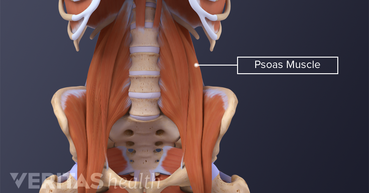
The Essential Role Of The Psoas Muscle from embed.widencdn.net There are also diseases and disorders that can cause the pain to. Your email address will not be published. Functionally, the hip joint enjoys a very high range of motion. The muscles that flex the hip are in front of the hip joint. The muscles of the neck can be divided into groups according to. Most modern anatomists define 17 of these muscles, although some additional muscles may sometimes be considered. Injury to the iliopsoas may cause hip pain and limited mobility. These muscles include the gluteus maximus muscle (the largest muscle in the body) and the hamstrings group, which consists of the biceps femoris, semimembranosus, and semitendinosus muscles.
Hip muscle anatomy is a complex topic.
Lateral rotation is needed for crossing the legs. Here we explain the hip and groin muscles, their actions and exercises. Iliopsoas muscle, a hip flexor muscle that attaches to the upper thigh bone. Related posts of muscles of the lower back and hip diagram muscle anatomy for massage. The gluteals make up the muscles of the buttocks on the back of the hip. Hip anatomy bones and joints of the hip. The hip joint is a ball and socket synovial joint, formed by an articulation between the pelvic acetabulum and the head of the femur. The iliofemoral, pubofemoral, and ischiofemoral ligaments represent the thickenings of the joint capsule. The femur may also rotate around its axis about 90 degrees at the hip. This mri hip joint axial cross sectional anatomy tool is absolutely free to use. The muscles of the neck can be divided into groups according to. One at the left hip, and one at the right hip. This muscle group also functions to keep the femur head trapped within the hip socket.
Your email address will not be published. The hip's essential muscles are the sartorius, rectus femoris, gluteus minimus and medius, iliopsoas, adductors, and hamstrings. The hamstrings are three muscles at the back of the thigh that affect hip and knee. Attached to the bones of the skeletal system are about 700 named. 4, obturator internus m this webpage.
Muscles Of The Hips And Thighs Human Anatomy And Physiology Lab Bsb 141 from s3-us-west-2.amazonaws.com The hip joint is a ball and socket synovial joint, formed by an articulation between the pelvic acetabulum and the head of the femur. The quadriceps group of four muscles. The muscular system consists of the skeletal muscles and their associated structures. Hip anatomy bones and joints of the hip. If soft tissue, such as skin, muscles, fat, and fascia get strained or injured, left hip pain can come from the abdominal wall. The hamstrings are three muscles at the back of the thigh that affect hip and knee. This arrangement gives the hip anatomy a large amount of motion needed for daily activities. Here we explain the hip and groin muscles, their actions and exercises.
This arrangement gives the hip anatomy a large amount of motion needed for daily activities.
These muscles include the gluteus maximus muscle (the largest muscle in the body) and the hamstrings group, which consists of the biceps femoris, semimembranosus, and semitendinosus muscles. The hip joint is a ball and socket synovial joint, formed by an articulation between the pelvic acetabulum and the head of the femur. The piriformis, hamstring and gluteal muscles are found on the buttocks, and the main extensor of the hip is the gluteus maximus. Related posts of muscles of the lower back and hip diagram muscle anatomy for massage. Attached to the bones of the skeletal system are about 700 named. Adductor muscles on the inside of your thigh. The iliofemoral, pubofemoral, and ischiofemoral ligaments represent the thickenings of the joint capsule. Anterior muscles extend your legs and flex your thighs. Iliopsoas muscle, a hip flexor muscle that attaches to the upper thigh bone. There are 3 main layers of hip abductor muscles: Trunk muscles, 289 muscles of the thorax, 289 muscles of the abdominal wall, 289. The right and left hip bones also converge anteriorly to attach to each other. Hip muscle anatomy is a complex topic.
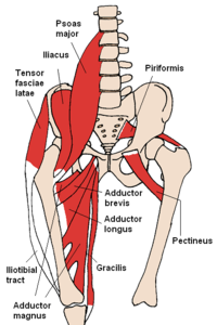
For detailed anatomy of pelvic bones, read anatomy of hip bone. The hip muscles are composed of multiple flexors, extensors, adductors, abductors, and rotators that work together. This muscle group also functions to keep the femur head trapped within the hip socket. Each hip bone, in turn, is firmly joined to the axial skeleton via its attachment to the sacrum of the vertebral column. Some of the other muscles in the hip are:
Muscles Of The Hip Wikipedia from upload.wikimedia.org Functionally, the hip joint enjoys a very high range of motion. They also stabilise the hip joint by 'pulling' the femoral head into the acetabulum of the pelvis. The hip's essential muscles are the sartorius, rectus femoris, gluteus minimus and medius, iliopsoas, adductors, and hamstrings. Anterior muscles extend your legs and flex your thighs. Use the mouse scroll wheel to move the images up and down alternatively use the tiny arrows (>>) on both side of the image to move the images.>>) on both side of the image to move the images. These bones are the thigh bone (femur) and pelvis. And what is trendelenburg's sign?daily anatomy app:for a random hum. The muscles of the neck can be divided into groups according to.
The strong muscles of the hip region also help to hold the hip joint together and prevent dislocation.
The hip muscles are composed of multiple flexors, extensors, adductors, abductors, and rotators that work together. These ligaments reinforce and stabilize the hip joint(6). Here we explain the hip and groin muscles, their actions and exercises. The hamstrings are three muscles at the back of the thigh that affect hip and knee. Attached to the bones of the skeletal system are about 700 named. These muscles are the adductor longus, adductor brevis, adductor magnus, gracilis, and the obturator externus. The muscles of the neck can be divided into groups according to. Trunk muscles, 289 muscles of the thorax, 289 muscles of the abdominal wall, 289. Injury to the iliopsoas may cause hip pain and limited mobility. The muscular system consists of the skeletal muscles and their associated structures. And what is trendelenburg's sign?daily anatomy app:for a random hum. Iliopsoas muscle, a hip flexor muscle that attaches to the upper thigh bone. The right and left hip bones also converge anteriorly to attach to each other.